Biomedical Imaging Research Unit

Annual image competition - category winners - 2024

The category winners and winner of the Hilary Holloway Prize for Best Image are shown below. The highly commended award winners can be viewed here. There was also one special mention award made this year - see it here.

Click on each image to see a larger version.

Visualisation category and trophy winner for 2024


V_2024_300px

Darven Murali Tharvan

Faculty of Science

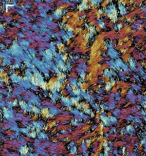
'When Van Gogh gets under your skin'

Reveals the organisation of collagen fibres in biological tissue showcasing the orientation and distribution of collagen fibres in human skin, taken in vivo.

The data was taken using polarisation-sensitive optical coherence tomography (PS-OCT).

In addition to receiving a mounted copy of his image and a bottle of wine, Darven receives a "basket of knowledge" to keep and the BIRU image trophy which he can keep for one year. His image will also decorate the BIRU home page for one year.
See Darven receiving his prizes from BIRU Technical Manager Richard Yulo.

Electron microscopy


EM_2024_300px

Morgan Liu

School of Chemical Sciences

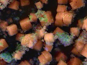
'Edible Carrot Cubes'

The samples are cyclodextrin-based metal-organic frameworks (MOFs) designed as carriers for metal-containing antimicrobials aimed at food preservation. The two clusters, differing in dimensions, illustrate different molecular interactions, the "LEGO"-Like assembly pattern, and the elemental distribution following chemical synthesis

Images captured at the RCSMS Engineering Department of the University of Auckland using the ZEISS GeminiSEM 460. The images are naturally colorized based on EDS results for elemental analysis.
See Morgan receiving her prize from BIRU Technical Manager Richard Yulo!

Light microscopy


LM_2024_300px

Meagan Chamberlain

Centre for Brain Research (CBR)

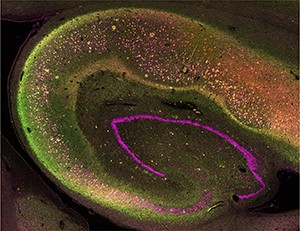
'Galaxy of Tau'

The beautiful expression of various phosphorylated tau species within the hippocampus of an Alzheimer's patient.

Imaged with the Olympus VS200 slide-scanner. 

See Meagan receiving her prizes from BIRU Technical Manager Richard Yulo.
Confocal microscopy


CLSM_2024_300px

Liam Zhang

Department of Physiology

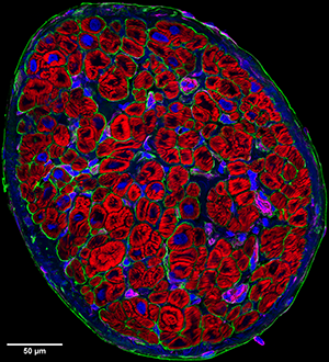
Human heart tissue

Imaged using the Zeiss LSM 800 confocal microscope.

It was captured using the Tile-scan feature with 63x magnification and processed using Huygens deconvolution.

See Liam receiving his prizes from BIRU Technical Manager Richard Yulo.