School of Medical Sciences


2009 competition - trophy and visualisation & analysis winner

Visualisation and analysis winner and trophy winner


va_chezviall_winner_160px

Chez Viall
Department of Obstetrics and Gynaecology
School of Medicine

This image shows first trimester placental villi that have been cultured for 24 hours. The cytoplasm of cells making up the villi are stained green with Cell Tracker CMFDA, and nuclei counter-stained with propidium iodide (here white). The z-stack of the villi was imaged at 20X magnification on the Zeiss LSM 710 inverted confocal microscope (BIRU) and the 3D rendering processed with Zen software.

Click on the thumbnail image to see a larger version.

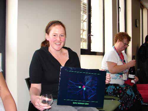
See Chez with her prize.

View another image that received a highly commended award

Light microscopy winner


lm_juliettecheyne_winner_160px

Juliette Cheyne
Department of Physiology, School of Medical Sciences

Fluorescence image of dissociated hippocampal cultures (21 days in vitro). Dendrites of neurons stained with microtubule associated protein 2 (MAP2, blue), Golgi stained with GM130 (red) and the granule cell marker calbindin (green).

Imaged at 63x magnification on the Zeiss Axioplan2 widefield fluorescence microscope

Click on the thumbnail image to see a larger version.

See Juliette with her prize.

View another fluorescence image that received a highly commended award

Electron microscopy winner


em_vijay_winner_160px

Vijayaraghavan Rajagopal
Auckland Bioengineering Institute

A longitudinal view inside a heart cell (acquired with a transmission electron microscope) using a new tissue preparation method that removes the grid-staining procedure. Involves HPF-FS.

Preparation and imaging work done in collaboration with Ying Jones at NCMIR (UCSD). This image shows that you don’t need grid-staining anymore.

Click on the thumbnail image to see a larger version.

See Vijay receiving his prize.

View another electron microscope image that received a highly commended award

Confocal laser scanning microscopy winner


clsm_cameronjohnson_winner_160px

Cameron Johnson,
Department of Opthalmology, School of Medicine

Section of Wistar rat retina following a partial optic nerve transection. Connexin43 (Alexa488; green) and glial fibrillary acidic protein (Cy3; red) label appears highly concentrated in the nerve fibre and ganglion cell layers. Cell nuclei are labelled with DAPI (blue). Images were collected at 60x magnification on the Ophthalmology Olympus FV1000 confocal microscope.

Unfortunately, Cameron was not available to collect his prize in person.

View another confocal image that received a highly commended award