Linda Graham & Jaswin Narayan
LabPLUS
Flat Stick - crystals within the renal podocytes composed of light chain
Acquired on the Tecnai G2 Spirit Twin transmission electron microscope
Click on thumbnail image to see larger version.
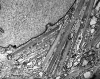
Flat Stick - crystals within the renal podocytes composed of light chain
Acquired on the Tecnai G2 Spirit Twin transmission electron microscope
Click on thumbnail image to see larger version.
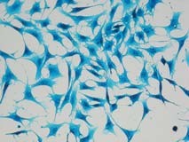
C17.2 cells grown in culture that have been fixed with 4% PFA and stained with blue food colouring
Equipment: 20x objective on a Nikon Eclipse TE2000-S microscope .
Click on thumbnail image to see larger version.
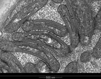
SAFETY PINS
Equipment BIRU Tecnai G2 Spirit TWIN electron microscope.
Click on thumbnail image to see larger version.
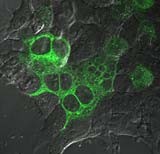
Live imaging of HEK293 cells expressing human melanocortin 4 receptor tagged with eGFP on Olympus FV1000 LCI microscope. Confocal images were overlaid with DIC images to observe rapid movement of vesicles containing hMC4R-eGFP
Imaged on the Olympus FV1000 Live Cell Imaging Confocal Microscope.
Click on thumbnail image to see larger version.
Download Emma's movie (10MB MPEG)
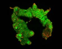
Placental Villus (or the Dying Caterpillar)
Three dimensional reconstruction of a single villus from a human placenta, with the cytoplasm stained green and nuclei stained red. Clusters of nuclei can be seen forming projections from the villus surface. These projections may be newly growing villi or represent the packaging of old nuclei. Click on thumbnail image to see larger version.
Now published - lyve1 expression reveals novel lymphatic vessels and new mechanisms for lymphatic vessel development in zebrafish.
Click on thumbnail image to see larger version.
Lateral image of Tg(lyve1:DsRed2;kdrl:EGFP) transgenic zebrafish line showing the blood vessels (green), lyve1 positive veins (yellow) and lymphatic vessels (red) at 12 days post fertilisation. The whole larva was imaged Equipment: Nikon D-Eclipse C1 confocal microscope (MMP).
lyve1 expression reveals novel lymphatic vessels and new mechanisms for lymphatic vessel development in zebrafish. Kazuhide S KS Okuda, Jonathan W JW Astin, June P JP Misa, Maria V MV Flores, Kathryn E KE Crosier, Philip S PS Crosier Development 139(13):2381-91 (2012). Kazuhide also managed to get another of his images on the journal cover!
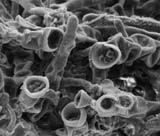
MICROSCOPIC BIRDS NEST FUNGI
Fruiting bodies of unidentified sooty mould fungi (from Kanuka infested with honeydew scale insect) with characteristic resemblance to ‘Bird’s nest fungi’
Equipment: ESEM FEI Quanta 200F with a SiLi (Lithium drifted) EDS detector Mag: 3000 X
Click on thumbnail image to see larger version.

