University home
parent of
Faculty of Medical and Health Sciences
parent of
School of Medical Sciences
parent of
ABOUT
parent of
Our departments
parent of
Biomedical Imaging Research Unit
parent of
Microscopy and imaging
parent of
Confocal microscopy
parent of
Olympus FV1000
Biomedical Imaging Research Unit
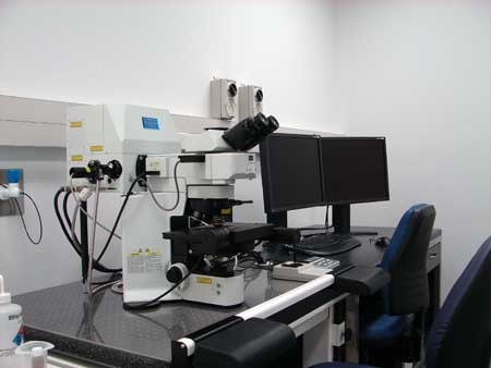
Olympus FV1000 confocal microscope - slide imaging
Specifications
- LASERS:
Diode (405nm, 440nm, 473nm, 635nm);
Diode-pumped (559nm) - Detectors: 2 spectral (variable bandpass), 1 filter-based, and one transmitted light
- Modes: xy, xyz, xt, xyt, lambda
- Spectral unmixing
- Resolution: up to 4096 x 4096 pixel resolution
- Fully motorised BX61 upright microscope
- Filter blocks for viewing specimens: U-MNU2 (narrow UV) U-MWBV (wideband blue) U-MNIBA3 (narrowband blue), U-MWIG3 (wideband green)
- Objectives: dry (4x/0.16 NA, 10x/0.40) and oil immersion (20x/0.80, 40x/1.00, 60x/1.35, 100x/ 1.40).
- Differential interference contrast available for all objective lenses
- Prior motorised XY stage, fully controllable through software
- Kinetic Systems anti-vibration table
Software
- FluoView 4.2
- Complete off-line licensed copy available in BIRU computer room for postprocessing
- Free FluoView viewer available for users. You can find it on the BIRU server or download it directly from the Olympus website here.
- Look here for other software packages available within the BIRU for processing confocal datasets
Funded by
School of Medical Sciences, Faculty of Medical and Health Sciences, The University of Auckland
Contact person
Jacqueline Ross
Lead Technologist, Technical Services
Email: jacqui.ross@auckland.ac.nz
Telephone: 923 7438 or Ext 87438
-
SCHOOLS, DEPARTMENTS AND CENTRES
Connect with us
Connect with us
-
SCHOOLS, DEPARTMENTS AND CENTRES


