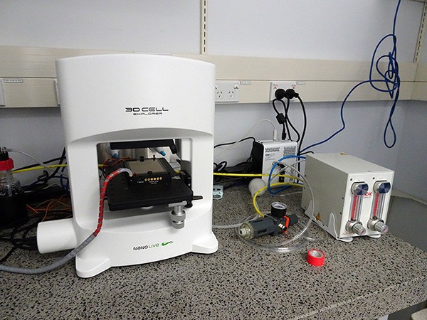
- 60x/0.8 NA objective lens, WD 300 microns
- High resolution – lateral 200nm, axial 400nm
- Field of view 85 x 85 x 30 microns
- Okolab incubation system - temperature and carbon dioxide control, compatible with microfluidics and perfusion systems
- Camera – CMOS, 165 frames/sec at 1024 x 1024 pixels
- Class I laser (520nm) low power
- One 3D reconstruction every 2 seconds
- Cool LED illumination source for fluorescence
- Triple-band filter set – FITC/TRITC/Cy5
University home
parent of
Faculty of Medical and Health Sciences
parent of
School of Medical Sciences
parent of
ABOUT
parent of
Our departments
parent of
Biomedical Imaging Research Unit
parent of
Microscopy and imaging
parent of
Holotomographic imaging
Biomedical Imaging Research Unit
Imaging capabilities
- 3D holotomographic imaging - label-free
- Fluorescence imaging - 2D only
- Quantitative phase analysis - calibration available
- Colour-coding (“painting”) based on refractive index
- Live cell imaging - up to 96 hours
Specifications

Specimen requirements
•Live or fixed cells grown on coverslips or in ibidi dishes (<30 microns thick)
•Tissue sections 5-10 microns thick unless very transparent
•Mounting media for tissue sections must be aqueous-based, e.g. Fluoromount, Prolong Gold
•Media for live imaging should be low scatter, wide range of media compatible with the system
•Samples cannot be dry
Documentation
Useful supporting materials for the system are available here including "how to" videos. Printed copies are also available in the microscope room.
Contact person
Jacqueline Ross
Lead Technologist, Technical Services
Email: jacqui.ross@auckland.ac.nz
Telephone: 923 7438 or Ext 87438
Connect with us
-
SCHOOLS, DEPARTMENTS AND CENTRES

