Biomedical Imaging Research Unit

Highly commended awards - 2014

Visualisation and Analysis - highly commended award


HC_David_VA_160px

David Crossman, Yufeng Hou, and Christian Soeller

Department of Physiology, School of Medical Sciences

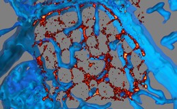
Correlative confocal and super resolution imaging of human cardiac myocyte

The visualisation shows a diseased human cardiac myocyte imaged with super resolution microscope for the protein junctophilin (in red) overlaid onto 3D confocal image stack of WGA labelling of the same cell (in blue). The blue tubular structures projecting into the centre of the cell are transverse tubules that are approximately 300 nm in diameter and are near the optical limit of the confocal microscope. However the super resolution data for junctophilin is substantially below this limit with a resolution of 30 nano-metres.


The image demonstrates how super-resolution and confocal microscopy can be combined to provide multi-scale imaging. Transverse tubules and junctophilin are key components in the calcium signalling that regulates cardiac contraction. Our research is characterising how changes in these structural components contribute to human heart failure.
 

Click on thumbnail image to see larger version

See David receiving the award!


Light microscopy - highly commended award


HC_IrinaRikus_Light_160px

Irina Rudykh, Rikus Botha

Department of Physiology, School of Medical Sciences

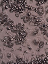
Lipid droplet formations in adipocytes 20x (fixed with  PFA and stained with Oil Red O)

Click on thumbnail image to see larger version

Light microscopy - highly commended award


HC_Zakieh_Light_160px

Zakieh Vahdati
Infection and Immunity, School of Medical Sciences

Supervisors Dr Siouxsie Wiles and Dr Simon Swift


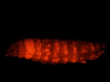
Galleria Mellonella larvae infected with fluorescently tagged M. marinum, ( day 8 post infection) lateral view

Acquired on Nikon SMZ1500 microscope

Click on thumbnail image to see larger version

Light microscopy - highly commended award


HC_Brad_Light_160px

Brad Raos
Department of Engineering Science, Faculty of Engineering

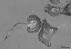
Reflective interference contrast image of live astrocytes in culture, captured at 60x on the Live Cell Olympus BX53 at the CBR

Click on thumbnail image to see larger version

See Brad receiving his award!

Confocal laser scanning microscopy - higly commended award


HC_Confocal_Pauline_160px

Pauline Misa
Crosier Laboratory, Department of Molecular Medicine & Pathology, School of Medical Sciences, FMHS

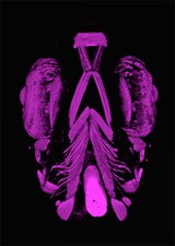
A swimming muscle-head
 
The ventral head musculature of a 5 day old zebrafish larvae, visualised by rhodamine-phalloidin labeling to reveal actin filaments of muscles. 
The image was acquired on a Nikon D-Eclipse C1 confocal microscope and 3d-rendered using Volocity.

Click on the thumbnail image to see a larger version

See Pauline with her prize