School of Medical Sciences


2008 competition -highly commended entries

Confocal laser scanning microscopy


caroline_highlycommended_clsm_160px

Caroline Brownline Brown
Microbial Ecology Lab, School of Biological Sciences

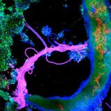
Maximum projection of an activated sludge sample. This sample was hybridised with DNA probes specific for all Bacteria (green) and members of the gamma-Proteobacteria (red), counterstained with DAPI (blue). It was visualised with the 63x oil immersion objective.

Imaged on the Leica TCS SP2 confocal microscope.

Light microscopy


vidya_highlycommended_lm_160px

Vidya WashingtonWashington
School of Biological Sciences

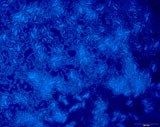
Serratia dry lower layer focused - DAPI Staining 

Juliette_highlycommended_160px

Juliette Cheyne
Department of Physiology

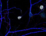
My first AMPA receptor! Fluorescence image of a newborn neuron in a dissociated hippocampal culture (13 days in vitro). New generated cells labelled with BrdU (white), dendrites of neurons stained with microtubule associated protein 2 (MAP2, Alexa 350, blue), synapses stained with synapsin-I (red) and the GluR1 subunit of AMPA receptor (green).

Imaged at 63x magnification on the Zeiss Axioplan2 widefield fluorescence microscope.

janie_highlycommended_lm_160px

Janie Foote
Department of Physiology

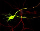
High molecular weight protein (Map2b-GFP, green) transfected neurons (shown by Map2, in red).

Imaged at 40x magnification on the Zeiss Axioplan2 widefield fluorescence microscope.

Visualisation and analysis


jeremy_highlycommended_va_160px

Jeremy Corfieldy Corfield
Department of Anatomy with Radiology/School of Biological Sciences

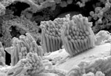
Kiwi haircells – an image of the stereovilli of a haircell in the kiwi ear. Imaged using a scanning electron microscope