School of Medical Sciences


BIRU image competition 2009 - highly commended entries

Confocal laser scanning microscopy


clsm_emma_kay_highlycommended_160px

Emma Kay
Department of Physiology

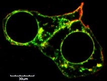
Single slice fixed confocal picture of HEK293 cells expressing the human melanocortin 3 receptor (green) and human melanocortin receptor 2 accessory protein (red).

Acquired on the Olympus FV1000 Live Cell Imaging system and image processed using ImageJ.

Click on thumbnail image to see larger version.

See Emma with her prize and others who received awards.

Light microscopy


lm_renee_gordon_highlycommended_160px

Renee Gordon
Department of Pharmacology and Clinical Pharmacology

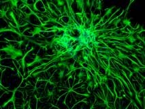
The image is x10 magnification taken on the Nikon Eclipse TE2000U microscope in the Connor lab. The cells are dividing neural stem cells from the adult rat brain which were allowed to differentiate into mature neural cells over 3 weeks in culture. The green staining is GFAP-positive astrocytes and the blue staining is DAPI stained nuclei.

Click on thumbnail image to see larger version.
See Renee leaving with her prize.

Electron microscopy


em_linda_graham_highlycommended_160px

Linda Graham
LabPLUS

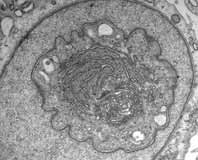
Nuclear pseudo inclusion. Golgi & RER seen through a window in a nucleus.

Imaged on the Tecnai G2 Spirit TWIN electron microscope.

Click on thumbnail image to see larger version.
See Linda with her prize.

Visualisation and analysis


va_sarah_rynbeck_highlycommended_160px

Sarah Rynbeck
Department of Physiology

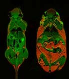
MRI images of lean C57BL6/J (left) and obese C57BL/6J (right) mice. Note the green area is anatomical (ie, non fat) while the orange area is fat. A series of images is acquired for each mouse and then used to work out the body composition of the mice (ie, percentage body fat).

Imaged on the high field MRI scanner using the Dixon method and processed using ImageJ.

Thanks go to Dr Beau Pontré for his assistance with the imaging.

Click on thumbnail image to see larger version.
See Sarah, Beau and other award winners.