Dedeepya Uppalapati
School of Pharmacy
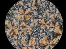
Liquid crystals under polarised light (20x)
Equipment: Leica DMR light microscope
Click on thumbnail image to see larger version.
Liquid crystals under polarised light (20x)
Equipment: Leica DMR light microscope
Click on thumbnail image to see larger version.
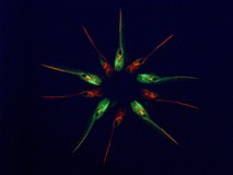
Fish meeting
Live imaging of Tg(lyve1:DsRed2;kdrl:EGFP) double transgenic zebrafish line and Tg(lyve1:DsRed2) single transgenic zebrafish line at 7 days post fertilisation. EGFP marks the blood vasculature while DsRed2 marks veins and lymphatic vessels.
Equipment: Leica MZ16 FA Fluorescence Stereomicroscope
Click on thumbnail image to see larger version.
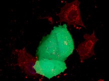
Three alive, three dead
3D-rendering of a z-stack taken through MCF-7 breast cancer cells treated with an anti-cancer agent delivered by a cell-penetrating peptide (red) and stained with Hoechst (blue) and cell-tracker (green).
Equipment: Zeiss LSM 710 confocal microscope
Click on thumbnail image to see larger version.
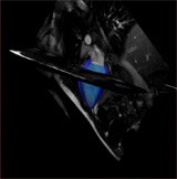
3D heart movement visualisation
Equipment: Siemens 3T MRI scanner
Click on thumbnail image to see larger version; watch movie
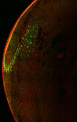
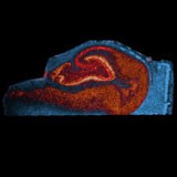
Lipids in the adult human hippocampus
This image was acquired using Matrix-Assisted Laser Desorption and Ionisation (MALDI) imaging. MALDI imaging enables one to visualise the distribution of any ion in the acquired mass spectrum, in the tissue of interest. This image was acquired at a 100 µm resolution, and the three different distributions were overlaid and pseudo-coloured using Photoshop.
Equipment: Applied Biosystems Voyager DE Pro MALDI-TOF; Adobe Photoshop
Click on thumbnail image to see larger version.
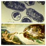
“Change and continuity”
Top image: thin sectioned UPEC 536 cell pellet visualised with 120Kv Tecnai G2 Spirit Twin TEM. Bottom image: “Creation of Adam”, Michelangelo Buonarroti, circa 1511.
Equipment: Tecnai G2 Spirit Twin transmission electron microscope
Click on thumbnail image to see larger version.

