Biomedical Imaging Research Unit

Image competition results 2016 - winners

The award winners were announced at the end of year Research Celebration on Thursday 8 December.

To view larger versions of the winning images, click on the smaller versions below.

Confocal microscopy


28 - salim Ismail -Combined spheres Proj

Himanshu Wadhwa, Salim Ismail, Jane McGhee and Trevor Sherwin

Department of Ophthalmology

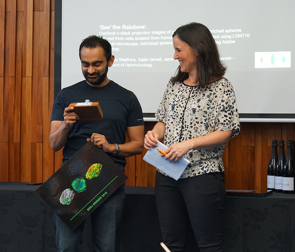
"See the Rainbow" - confocal z-stack projecion images of corneal stem cell-enriched spheres cultured from cells isolated from human ocular tissue.

Imaged using Zeiss LSM 710 confocal microscope, individual sphere images combined using Adobe Photoshop.

Sadly, Himanshu wasn't able to accept his prize in person but it was received by his colleague Salim who also contributed to the image.

See Salim receiving the trophy from Sue on Himanshu's behalf

See Himanshu with his supervisor Associate Professor Trevor Sherwin

Light microscopy


15 - reprogrammed human neurons K Jones-200px

Kathryn Jones – Centre for Brain Research

Beautiful neurons reprogrammed fluorescing to show TuJ1 (green) and (TH) a dopamine marker in red, along with DAPI

See Kat with her prize
 

Visualisation and analysis


YeLi 4 stills (002)-200px

Ye Li, Salim Ismail, Jane McGhee and Trevor Sherwin
Department of Ophthalmology

"Fungal Invasion" - Time-lapse microscopy of a stem cell sphere cultured from a cadaveric cornea with keratoconus and placed on collagen. Imaged on Nikon Biostation.

See Ye wih her prize

Electron microscopy - scanning electron microscopy


02 -Josh Workman SEM Cartilage-200px

Josh Workman -
Scanning Electron Microscopy (SEM) image of fracture surface in articular cartilage surface. Taken using an FEI (Phillips) XL30 S-FEG

See Josh with his prize
 

Electron microscopy - transmission electron microscopy


23 - LindaG -image_40-200px

Linda Graham - LabPlus

Renal biopsy

See Linda with her prize