Linda Graham & Jaswin Narayan
LabPLUS
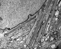
Flat Stick - crystals within the renal podocytes composed of light chain
Acquired on the Tecnai G2 Spirit Twin transmission electron microscope
Click on thumbnail image to see larger version.
Flat Stick - crystals within the renal podocytes composed of light chain
Acquired on the Tecnai G2 Spirit Twin transmission electron microscope
Click on thumbnail image to see larger version.
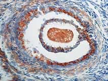
Tethered by a thin stalk of zona pellucida cells, an oocyte floats within its follicle.
Equipment: Taken with the Nikon Eclipse E400.
Click on thumbnail image to see larger version.
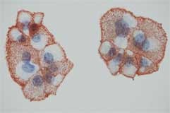
Dead or alive? Aggregations of nuclei shed from a human placenta blush pink where damaged DNA is present.
Equipment: composite of several images taken with the Olympus FV1000 confocal microscope
Click on thumbnail image to see larger version.
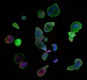
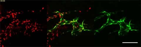
Imaged on the Olympus FV1000 Live Cell Imaging Confocal Microscope.
Live imaging of the zebrafish facial lymphatic vessel and pectoral fin vessel at 1.5 to 2 days post fertilisation. lyve1 expression (green) shows vessel development and nuclear kdrl expression (red) shows the single cell migration. Scale bar: 100 µm. Time: 9 hours
Click on thumbnail image to see larger version.
See Kaz's movie (6MG GIF)

