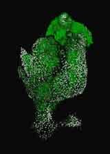
The winning image shows first trimester placental villi that have been cultured for 24 hours. The cytoplasm of cells making up the villi are stained green with Cell Tracker CMFDA, and nuclei counter-stained with propidium iodide (here white).
The z-stack of the villi was imaged at 20X magnification on the Zeiss LSM 710 inverted confocal microscope (BIRU) and the 3D rendering processed with ZEN software.
Click on thumbnail image to see a larger version.


