Biomedical Imaging Research Unit

Highly commended awards - 2024

The images below all received highly commended awards.

Visualisation


HC_V_2024_300px_R

Christian John Saludar, Maryam Tayebi, Eryn Kwon, Vickie Shim

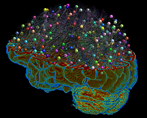
'Wired Neural Wonders'

A depiction of the astounding complexity yet intricate beauty of the brain. The piece illustrates the power of tractography, an advanced medical imaging technique that allows visualization of fiber tracts composed of billions of NEURONS,  connecting brain regions resembling a wonderful WIRED circuitry of interconnected nodes. 

Complex as it may seem, with all the varying components, the brain's total beauty and significant functionality leave anyone in awe and WONDER.

This multi-shell diffusion MR image was taken using a GE 3.0 T SIGNA Premier MRI scanner at Matai Medical Research Institute.

See Christian receiving his prize from BIRU Technical Manager Richard Yulo.

Electron microscopy


HC_EM_2024_300px

Al Gerard Des Los Reyes Makilan

LabPlus

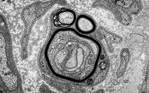
'Nerve-ous Scream'

A regenerating myelinated axon (nerve tissue).

Acquired on the FEI Tecnai Spirit TWIN transmission electron microscope.

See Al receiving his prize from BIRU Technical Manager Richard Yulo.
Light microscopy


HC_LM_2024_300px

Stanley Cardon

Centre for Brain Research

Microscope used: Olympus Slideview VS200 slide-scanner

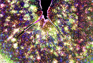
The image was achieved using a form of fluorescent immunohistochemistry called multiplex immunohistochemistry where multiple rounds of immunohistochemistry can be performed on a single piece of tissue by stripping antibodies between rounds. The image is taken from a section from the temporal-frontal gyrus of an individual with Alzheimer's disease where we have labelled the below listed proteins using 5 different antibodies with different fluorophores and colours.
 

These antibodies have almost never before been used to label these proteins in post-mortem brain tissue in Alzheimer's disease before and from the image we can see that these proteins are all labelling astrocytes in the grey matter, all with different states and functions which are accumulating around the vast distribution of beta-amyloid plaques in a sort of "astrocytic firework" of colour. 

Green = Aquaporin 1 , Magenta = CD99 , Red = GFAP , Blue = Aquaporin 4, Yellow = SERPIN-A3

See Stanley receiving his prize from BIRU Technical Manager Richard Yulo.
Confocal microscopy


CLSM_HC_300px

Amelie Back

Department of Pharmacology and Clinical Pharmacology

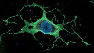
'Keeping an eye open'

A human-reprogrammed oligodendrocyte stained with the marker O1 (green).

See Amelie receiving her prize from BIRU Technical Manager Richard Yulo.