School of Medical Sciences


2006 competition - highly commended entries

Confocal laser scanning microscopy


moonlight-on-the-ocean_160px

"Moonlight on the ocean"
Elena Vazey
Department of Pharmacology

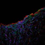
HCC projection confocal stack of GFAP positive rat glial cells along the edge of the cortex and within a human embryonic stem cell derived neural progenitor cell graft 4 weeks after transplantation into excitotoxically lesioned rat brain graft not shown. The image was taken at 40x mag on the BIRU Leica SP2. The slice distance was 0.35 um and the total stack covered a total z distance of 25.55 um. Dako rabbit anti GFAP 1:1000 with an Alexa fluor 594 conjugated anti rabbit secondary (1:500) was used. 

kiwifruit_highlyrecommended_160px

Lin-Chien Huang
Department of Physiology

Image now published in: Huang LC, Thorne PR, Housley GD, Montgomery JM. Spatiotemporal definition of neurite outgrowth, refinement and retraction in the developing mouse cochlea. Development. 2007;134:2925-33. Epub 2007.

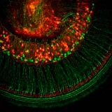
Confocal image of a whole-mount preparation of postnatal day 6 mouse cochlea, labelled with tetramethylrhodamine-conjugated dextran (red) and peripherin immunofluorescence (green) to represent the mature primary afferent innervation. This image clearly demonstrates an independent labelling of two different markers on a stripe of primary auditory neurones and their peripheral nerve fibres projected radially towards the sensory hair cells. The innervation pattern of the primary auditory neurones and their projections resembles a Kiwi fruit slice. This image was taken on the BIRU Leica TCS SP2. 

soeller_gilbert_highlycomm_160px

Dr Christian Soeller & Ray Gilbert
Department of Physiology

Image now published in: Christian Soeller, David Crossman, Ray Gilbert and Mark B. Cannell, Analysis of ryanodine receptor clusters in rat and human cardiac myocytes, PNAS, September 18, 2007, 104; (38), 14958-14963.

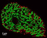
Cross-section through an isolated rat ventricular cardiomyocyte, stained for both alpha actinin (green) and RyR2 receptor (red). The green patches correspond to bundles of contractile machinery and the red puncta represent clusters of ryanodine receptors that release calcium from the sarcoplasmic reticulum upon activation. Cells were orientated end on using a novel agar embedding method, allowing unprecedented resolution in cross section (~250 nm), that is largely unaffected by the blurring artefacts normally resulting from the limited Z resolution of confocal microscopy. Note the arrangement of release clusters around the contractile machinery which is activated by the calcium rapidly diffusing from the site of release.

Image captured on a Zeiss LSM410 confocal microscope in the Physiology Dept. FMHS and processed using custom written deconvolution software.

Sample preparation and imaging: Ray Gilbert
Data processing and display: Christian Soeller
Funding: Auckland Medical Research Foundation 

Light microscopy


lm_highlyreccomended_160px

Claire French
Department of Anatomy with Radiology

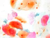
Brightfield image of epithelial cells from inside the mouth. It illustrates the differential expression of keratin within the oral cavity. The image was captured on the Leica DMR light microscope at 63x.  

hippocampus_jbai_160px

Dr Ji-Zhong Bai
Department of Physiology

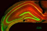
The image shows the structure of rat hippocampus, stained green with Nissl Green for neuronal cells and red with immuno-staining of Transient Receptor Potential V4 (TRPV) channels. This image was captured with a Zeiss Fluorescence Microscope with standard FITC and Rhodamine filters using 4x objective.  

Medical imaging


medical_imaging02mouse-tibi_160px

Garry Williams
Department of Medicine

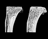
3D model of mouse tibiae. Tibiae were scanned by micro computed tomography at a 5um cubic voxel resolution, using the Skyscan 1172 desktop scanner, located in the Bioengineering Institute. The bone on the left is control, the bone on the right is from an adiponectin deficient animal.