Biomedical Imaging Research Unit

Highly commended awards - 2021

Highly Commended awards are given where the image or video has particular merit and just misses out on being the winner of a category. 

Please click on the image to see a larger version...

Visualisation and analysis


VA_HC_300px2

Anna Krstic

Department of Physiology

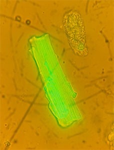
Calcium transients from a contracting cardiomyocyte

This video (see below) shows the way calcium is released on a beat-to-beat basis in a single myocyte out of millions that contribute to contraction of the whole heart.

It was taken down the eyepiece of the Zeiss LSM 710 confocal microscope.

See Anna after receiving her prize

Electron microscopy


EM_HC_300px

Teena Gamage

Department of Physiology

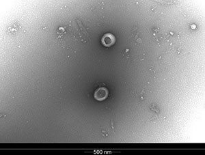
Fetal sheep plasma-derived extracellular vesicles

See Teena with her prize

Light microscopy


LM_HC_300px

Shaima Albeloushi

Liggins Institute

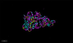
Islet of Langerhans in sheep. Insulin Beta-cell (yellow), Glucagon alpha-cells (pink), and Somatostatin delta-cells (blue)

See Shaima after receiving her prize

Confocal (super-resolution) microscopy


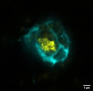
This image was entered under the Confocal Microscopy category however it is actually a super-resolution image acquired on the BIRU's newest system, the Abberior Facility Line Stimulated Emission Depletion (STED) microscope. We look forward to seeing more entries acquired on this system in 2022.

CLSM_HC_300px

Victor Dieriks,

Anatomy & Medical Imaging, CBR

'Blue and Yellow Nebula'

Breaking through optical resolution barriers. Unraveling layered aggregate pathology in the human brain. The image was acquired on the Abberior STED microscope.

See Victor with his prize.