Dr Henry Waldvogel & Ray Gilbert
Department of Anatomy with Radiology
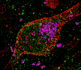
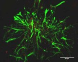
Neuron in the human brain.
Tissue preparation (Henry Waldvogel)
Imaging, deconvolution & 3D reconstruction (Ray Gilbert).
Imaged on the Leica TCS SP2 confocal microscope.
-
Biomedical Imaging Research Unit
- Our unit
- Microscopy and imaging
- Tissue processing
- Flow cytometry
- Image processing and analysis
-
BIRU image competition
- 2014 trophy winner
- 2013 trophy winner
- 2013 winners
- 2013 commended
- 2012 trophy winner
- 2012 winners
- 2012 commended
- 2012 special award
- 2011 trophy winner
- 2011 winners
- 2011 commended
- 2011 special awards
- 2010 trophy winner
- 2010 winners
- 2010 commended
- 2009 trophy winner
- 2009 winners
- 2009 commended
- 2008 trophy winner
- 2008 winners
- 2008 commended
- 2007 winners
- 2007 commended
- 2006 winners
- 2006 commended
- 2014 winners
- 2014 commended
- 2015 winners
- 2015 commended
- 2015 trophy winner
- 2015 special mention
- 2016 winners
- 2016 commended
- 2017 winnners
- 2017 commended
- 2018 commended
- 2018 winners
- 2019 winners
- 2019 commended
- 2019 trophy winner
- 2020 winners
- 2020 commended
- 2020 trophy winner
- 2021 trophy winner
- 2021 winners
- 2021 commended
- 2021 special award
- 2022 winners
- 2022 commended
- 2022 trophy winner
- 2022 special awards
- 2023 winners
- 2023 commended
- 2024 winners
- 2024 commended
- 2024 special mention
- 2025 winners
- 2025 commended
- Event photos
- Resources and links
- Teaching and learning
- Our people
- Equipment charges
- Seminars and workshops
University home
parent of
Faculty of Medical and Health Sciences
parent of
School of Medical Sciences
parent of
ABOUT
parent of
Our departments
parent of
Biomedical Imaging Research Unit
parent of
BIRU image competition
parent of
2007 commended
School of Medical Sciences
2007 competition -highly commended
-
Biomedical Imaging Research Unit
- Our unit
- Microscopy and imaging
- Tissue processing
- Flow cytometry
- Image processing and analysis
-
BIRU image competition
- 2014 trophy winner
- 2013 trophy winner
- 2013 winners
- 2013 commended
- 2012 trophy winner
- 2012 winners
- 2012 commended
- 2012 special award
- 2011 trophy winner
- 2011 winners
- 2011 commended
- 2011 special awards
- 2010 trophy winner
- 2010 winners
- 2010 commended
- 2009 trophy winner
- 2009 winners
- 2009 commended
- 2008 trophy winner
- 2008 winners
- 2008 commended
- 2007 winners
- 2007 commended
- 2006 winners
- 2006 commended
- 2014 winners
- 2014 commended
- 2015 winners
- 2015 commended
- 2015 trophy winner
- 2015 special mention
- 2016 winners
- 2016 commended
- 2017 winnners
- 2017 commended
- 2018 commended
- 2018 winners
- 2019 winners
- 2019 commended
- 2019 trophy winner
- 2020 winners
- 2020 commended
- 2020 trophy winner
- 2021 trophy winner
- 2021 winners
- 2021 commended
- 2021 special award
- 2022 winners
- 2022 commended
- 2022 trophy winner
- 2022 special awards
- 2023 winners
- 2023 commended
- 2024 winners
- 2024 commended
- 2024 special mention
- 2025 winners
- 2025 commended
- Event photos
- Resources and links
- Teaching and learning
- Our people
- Equipment charges
- Seminars and workshops
-
SCHOOLS, DEPARTMENTS AND CENTRES
Connect with us
Connect with us
-
SCHOOLS, DEPARTMENTS AND CENTRES


