Biomedical Imaging Research Unit

Image competition results 2017 - winners

The award winners were announced at the end of year Research Celebration on Thursday 23 November.

To view larger versions of the winning images, click on the smaller versions below.

Light microscopy category & trophy winner for 2017


CharlieJohnson_Light_small

Dr Charlotte Johnson

School of Biological Sciences

"Ascension": Natural fluorescence of a microsporophyll of a male pine cone captured at x 60 magnification using a Leica DMRE light microscope. Multiple focal planes were combined in Photoshop.

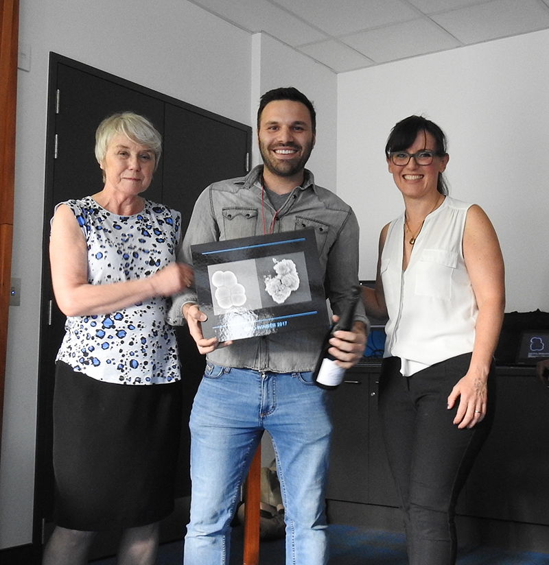
See Charlotte receiving her award for Light microscopy...

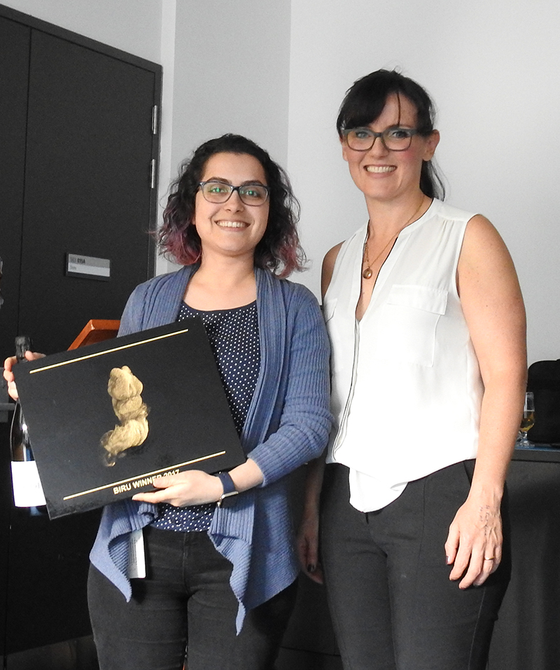
See Charlotte receiving the BIRU image trophy from BIRU Director Dr Sue McGlashan!

Electron microscopy


KyriakosVarnava_small

Kyriakos Varnava

School of Chemical Sciences

Staphylococcus aureus cells. Before(left) and after (right) treatment with the peptide

 Imaged using a Hitachi SU-70 FESEM, individual images combined using Adobe Photoshop.

See Kyriakos receiving his award from BIRU Director Dr Sue McGlashan!

Confocal microscopy


FrazerCoutinho_small

Frazer Coutinho

Department of Ophthalmology

‘Peacock Feathers in Full Bloom’

C57BL/6 mouse retinal section labelled with secondary Alexa Fluor 488 antibody, GFAP-Cy3 and DAPI nuclear stain. The image resembles a peacock showing off its feathers in full bloom. Acquired on, Olympus BX-61 confocal microscope (Department of Ophthalmology).

Visualisation and analysis


Naz_small

Nazanin Ebrahimi, Dane Gerneke, Mahyar Osanlouy

Auckland Bioengineering Institute

Alien embryo - a reconstructed micro CT from a 10 day old rat embryo. The embryo length is 1.5 mm. The image was acquired using a Bruker Skyscan 1272 at 0.45 micro meter resolution. From the video, we can see the single cells of the forming endocardium.

See Nazanin receiving her award from BIRU Director Dr Sue McGlashan!