Biomedical Imaging Research Unit

Highly commended awards - 2023

Highly Commended awards are given where the image or video has particular merit and just misses out on being the winner of a category. 

Click on each image to see a larger version or a movie if it is a video file entry.

Visualisation and analysis


VA_2023-HC2_movie_450px

Keon Manzanero and Aaron Chester

MEDSCI 300 students

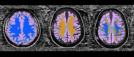
'Matter over mind'

Long-term methamphetamine use can permanently damage the brain, but it can recover to a certain extent: it can cause grey matter atrophy, shown in clear perspex.

Video shows segments created from visualisation software Amira. MRI scans used for visualisation obtained from the Mātai Medical Research Institute.
 

Left: Healthy, age-matched control

Centre: Chronic methamphetamine use

Right: 8 months after quitting methamphetamine use

Click on the image to see the video.

See Keon with Caleb Paterson
 

Visualisation and analysis


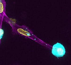
VA_2023-HC_movie

Evert Loef

School of Biological Sciences

Human monocyte-derive dendritic cell endocytoses a dead melanoma cell, one tiny bit at a time.
Video captured over 30 minutes on the Nikon Ti-e at the School of Biological Sciences using the spinning disk confocal system

Electron microscopy


EM_2023_HC_300px

Grace Casabuena

LabPlus

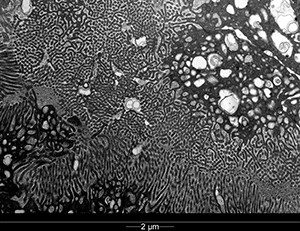
This is a high magnification electron micrograph of a renal biopsy from a patient with suspected Lupus Nephritis. The image shows proximal tubule with apical cytoplasmic regions and brush borders. Beneath the surface microvilli lies coated vesicles, apical dense tubules, and endosomes which are components of endocytic-vacuolar apparatus.

Image acquired on a FEI Tecnai Spirit TWIN transmission electron microscope.

Electron microscopy


EM_2023_HC2_300px

Pang Yuk Cheung

Department of Molecular Medicine & Pathology

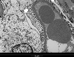
'Footsies'

Transmission electron microscope image of the podocyte foot processes from a rat kidney.

Image acquired on the FEI Tecnai Spirit TWIN transmission electron microscope.
 

Confocal microscopy


CLSM_2023_HC_300px

Janneke Grundermann

Auckland Cancer Society Research Centre

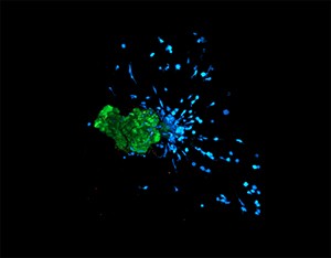
'3D organoid-macrophage co-culture' 
 
Mouse macrophages (blue) infiltrating into the matrigel droplet and interacting with mouse tumour organoids (green).
 
Macrophages stained with Cell Proliferation Dye eFluor 450, organoids stained with CellTracker Green Dye, and Propidium Iodide (red) used as cell death marker.

Imaged on the Zeiss LSM 800 Airyscan confocal microscope.
 

Light microscopy


LM_2023_HC_300px

Po-Yi Lue

Department of Physiology

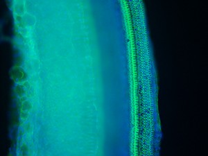
Sheep cochlear hair cells: this image is taken from the middle turn of a sheep cochlear wholemount. One row of inner hair cell and three row of outer hair cell can be identified. Also, some spontaneous hair cell loss can be observed.

Green: Phalloidin; Blue: DAPI.

Imaged on a Leica DMR fluorescence microscope