Biomedical Imaging Research Unit

Highly commended and special awards - 2018

The following awards were presented at the BIRU end of year Research Celebration on Thursday 29 November 2018. Congratulations to all of those who received awards and thanks to all who participated this year.

Confocal microscopy - highly commended

clsm_hc_vd_200px

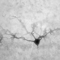
Victor Birger Dieriks, PhD

Department of Anatomy and Medical Imaging, Centre for Brain Research

A mitral cell in the olfactory bulb

Confocal microscopy - highly commended


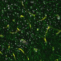
Fairy Lights in the Bamboo Forest

Karan Govindpani

Centre for Brain Research

Department of Anatomy and Medical Imaging

“Fairy Lights in the Bamboo Forest”

Cerebral blood vessels (yellow-green) and astrocyte processes (bright green) in the human middle temporal cortex, reminiscent of bamboo stems and leaves. Cell nuclei labelled with Hoechst stain in white. Taken at 20x magnification using the LSM710 laser-scanning confocal microscope


Confocal microscopy - special mention


clsm_sm_vs_250px

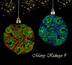
Veronika Sander

Department of Molecular Medicine and Pathlogy

The images are kidney organoids grown from human induced pluripotent stem cells, stained with beta catenin (green), PCNA (red) and Hoechst, taken on the 710 Zeiss confocal.

Confocal microscopy - special mention


clsm_sm_lm_200px

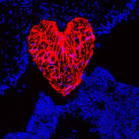
Lola Mugisho

Department of Ophthalmology

Eye’ love research

Confocal image showing glial fibrillary acidic protein (GFAP, red) labelling of the optic nerve in cross section. Cell nuclei stained with DAPI (blue). Image captured with a 20x objective lens on a Olympus FV1000 confocal laser scanning microscope


Electron microscopy - highly commended


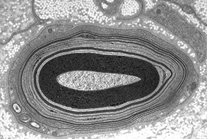
em_hc_lg_300px

Linda M Graham

Anatomical Pathology, LabPlus

“The nerve of it”

Image of an abnormal  myelinated nerve

Electron microscopy - highly commended


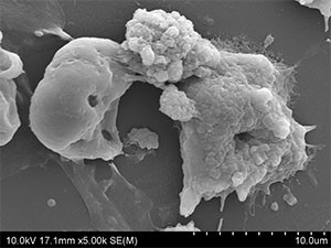
em_hc_kp_300px


Kamal Patel & Dr Viji Sarojini

School of Chemical Sciences

Faculty of Science

SEM image of treated and untreated Neuroblastoma cancer cells


Visualisation and analysis - highly commended


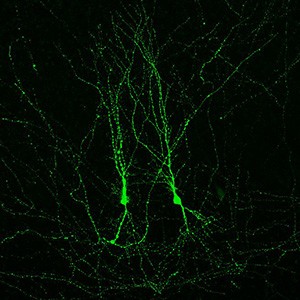
va_hc_yv_200px

Yukti Vyas

Centre for Brain Research, Department of Physiology

Movie showing Z-stack of hippocampal neurons from organotypic slice cultures transfected via electroporation with green fluorescent protein (GFP). 

Visualisation and analysis - highly commended


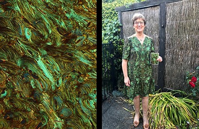
va_sm_swt_bone_model200px-jpg

Sharon Waldvogel-Thurlow and Nikki Mills

Department of Surgery

Taken on the DMR under polarised light. I helped Nikki Mills with the photography and then I had it printed into material and made this dress!

Light microscopy - highly commended


lm_hc_bh_200px

Blake Highet

Department of Anatomy and Medical Imaging, Centre for Brain Research

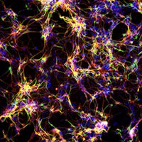
Mouse carcinoma P19 cells which  have been differentiated into neurons

Taken on ImageXpress

Light microscopy - highly commended


lm_hc_hm_200px

Helen Murray, PhD

Department of Anatomy and Medical Imaging, Centre for Brain Research

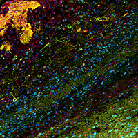
Sagittal human olfactory bulb section labelled using fluorescent multiplex IHC to illustrating the different neuronal subtypes types.

Markers:

DAPI (blue); NeuN (cyan); PGP9.5 (green); Parvalbumin (yellow); Calretinin (orange); Calbindin (red)