Biomedical Imaging Research Unit

Highly commended and special awards - 2019

The following awards were presented at the BIRU end of year Research Celebration on Wednesday 27 November 2019. Congratulations to all of those who received awards and thanks to all who participated this year.

Click on the small image to see a larger image or a  movie...

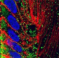
Confocal microscopy - highly commended


clsm_hc_2019_200px

Nandini Bavana

Dept of Physiology

Distribution of Aquaporin 1 water channel in the rat lens epithelium


Imaged on the BIRU Olympus FV1000 BX61 confocal microscope

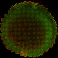
Confocal microscopy - special mention


clsm_sm_2019_200px

Nandini Bavana

Dept of Physiology

A multi area stitched image of rat lens axial section

During multi area scan imaging when the image is left on an angle a beautiful image came out from an error... 

Imaged on the BIRU Olympus FV1000 BX61 confocal microscope
 

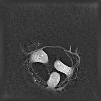
Visualisation and analysis - highly commended


va_hc_2019_200px

 Molly Abraham

Department of Physiology

“Circle of friendship”
 
Live unstained image of cultured hippocampal neurons taken with the Nanolive 3D Cell Explorer at 60x magnification
 

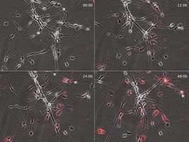
Visualisation and analysis - highly commended


va_hc_2019_2_200px

Victor Dieriks

Dept. Anatomy and Medical Imaging - Centre for Brain Research

“Hungry Boys”

Imaged on the Nikon BioStation live cell imaging system

Click on the image to see the video..

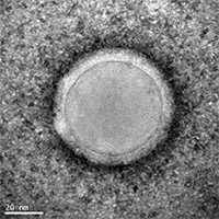
Electron microscopy - highly commended


em_hc_2019_200px

Denis Simonov

School of Biological Sciences & Department of Surgery

A human lymph exosome

Negative staining

Imaged using the FEI Tecnai T12 transmission electron microscope - magnifcation x150,000
 

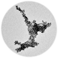
Electron microscopy - special mention - Patriot award


em_sm_patriot_2019_200px

Hugh Glossop

School of Chemical Sciences

Nano-North Island 

Gold nano-particles and peptides aggregated into a familiar shape

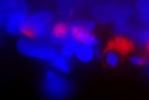
Light microscopy - special mention - Patriot award


lm_sm_patriot_2019_200px

Judy Loh, Jane McGhee and Trevor Sherwin

Department of Ophthalmology

‘Which came first, the kiwi or the egg?’

Immunolabelling of a diseased mucosal sample with C3B9 (red) and DAPI stain