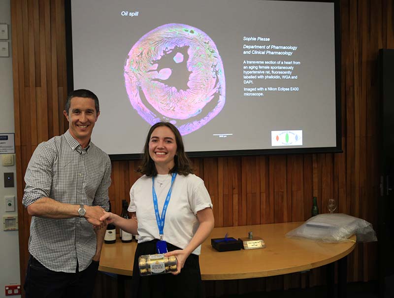
Biomedical Imaging Research Unit

Highly commended awards - 2022

Highly Commended awards are given where the image or video has particular merit and just misses out on being the winner of a category. 

Click on each image to see a larger version.

Visualisation and analysis


Frederique Vanholsbeeck, Ashly Jose, Department of Physics, Faculty of Science

Juliette Cheyne, Pang Ying Cheung, Zahra Laouby, Department of Physiology, School of Medical Sciences, FMHS

'Rat brain gets lit'

In-vivo imaging of the brain allows investigation of neural niches impacted by assaults on the central nervous system.  The time-lapse video displays the spontaneous neural firing of a rat's brain imaged through its skull.  The fluorescence images are captured using a custom-built and cost-effective (<10000 USD) mesoscope, a large field of view widefield microscope.

See Ashly with her prize

Video - 'Rat brain gets lit'

Light microscopy


LM_HC_300px

Sophie Piesse

Department of Physiology, School of Medical Sciences, FMHS

'Oil spill'

A transverse section of a heart from an aging female spontaneously hypertensive rat, fluorescently labelled with phalloidin, WGA and DAPI.

Imaged with a Nikon Eclipse E400 microscope.

See Sophie with her prize

Electron microscopy


EM_HC 300px

Laurenz Boettger

School of Environment, Faculty of Science

'Uvigerina peregrina (benthic foraminifera)'

Intact whole shell

Imaged on the Hitachi TM3030 Plus SEM at 600x magnification

See Laurenz's image at the presentation

See Laurenz with his prize

Confocal microscopy


CLSM_HC2_300px

James Wiseman

Department of Anatomy and Medical Imaging/CBR, School of Medical Sciences, FMHS

'α-synuclein aggregates in multiple system atrophy'

Each label detects different epitopes of the α-synuclein protein with the nuclei stained with Hoechst (blue).

Imaged at 63x with the Airyscan module on the Zeiss LSM 800 Airyscan confocal microscope

See James receiving his prize

Visualisation and analysis


Miriam_300px

Miriam Scadeng

Department of Anatomy and Medical Imaging, Faculty of Medical and Health Sciences

'Arapaima'

Arapaima are the world’s largest air breathing fish, reaching 2.5m in length, and live in piranha-rich Amazonian waters.


Upper image: Transparent surface rendering of Arapaima with internal organs as coloured surfaces.

Lower 3 images are lateral, superior and inferior views of protective skull and head

bones.

See Miriam's image at the Research Celebration