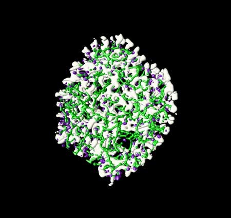
A slice through a cardiac muscle cell (one frame from a movie)
Isuru D. Jayasinghe, Christian Soeller
Red: contractile apparatus: F-actin (Alexa 594- phalloidin) Blue: predicted arrangement of mitochondria Grey: Z-disks anchoring actin filaments: (intensity-based segmentation of actin images)
Purple: Ca2+ release sites (Ryanodine receptors labeled via mouse anti- RyR2 / Atto-647) Cyan: transverse-axial tubular system (membrane marker caveolin-3 labeled via rabbit anti-cav3 / Alexa 488) White: surface membrane (spatial segmentation of caveolin-3)
Sample: Isolated and fixed rat ventricular myocyte
Imaging: Olympus FV1000 Live Cell Imaging Confocal System (BIRU)
Image processing: deconvolved using custom-written Richardson-Lucy maximum-likelihood algorithm, custom-written segmentation and tracing algorithms implemented in IDL 7.0 (ITT)
Surface rendering performed in Open DX (open source)
Movie generated using ImageJ AVI output, VirtualDub compression and editing performed with Windows Moviemaker 5.1; segmented data is used as input to construct cardiac calcium handling models with realistic geometry. The entire movie will be uploaded as soon as possible.


