Biomedical Imaging Research Unit

Image competition results - 2022

The award winners of the 2022 BIRU image competition were announced on Thursday 24 November at the annual BIRU Research Celebration. This year, we were able to hold the event in person. The BIRU Director, Dr David Crossman, was not able to be present in person but provided his speech as a video file.

The winner of each category received a mounted copy of their image (high quality print mounted on ArtMount), a bottle of Goldie sparkling chardonnay or a box of chocolates according to their preference. Those who received Highly Commended, Special Mention or Patriot awards received a bottle of Goldie wine or a box of chocolates. The prizes were presented by former BIRU Director, Dr Gus Grey with assistance from new BIRU staff members Dr Sandy Chen and Yohanes Nursalim.

The category winners are shown below. The Highly Commended and Special Mention and Patriot awards can be accessed using the links below. 

Click on each image to see a larger version.

See Highly Commended award winners

See Special Mention award and Patriot award winners

Light microscopy and trophy winner for 2022


Kyrah_300px

Kyrah Thumbadoo

School of Biological Sciences, Faculty of Science

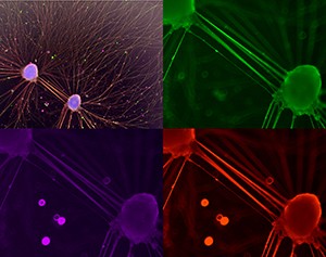
'Human connection'

iPSC-derived motor neurons successfully differentiated from fibroblasts of a person with Charcot-Marie Tooth disease, a peripheral neuropathy.

Specifically, two neurospheres form robust links to one another, and outward, creating highly connective axonal branches, as marked by neuron-specific cytoskeletal elements βIII-tubulin (red), and neurofilaments (green, magenta).

Imaged on the EVOS FL Auto 2 at 20x magnification in channels 488, 594, and 647

See Kyrah with the Hilary Holloway Prize for best image

Confocal microscopy


Dayna_300px

Dayna Spurling

Department of Pharmacology and Clinical Pharmacology, School of Medical Sciences, FMHS

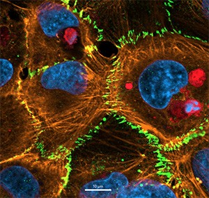
'Melanoma derived vesicles inside a single hCMVEC'

Uptake of melanoma derived vesicular bodies (red – CTR) by human brain endothelial cells following a 4 hour incubation.

Blue – nucleus (DAPI); Red – vesicular bodies (CTR); Orange – F-actin (ReadyProbes Actin AF 555); Green - Junctional CD144 (AF488)

Imaged at 40x oil immersion on the Zeiss LSM 800 confocal microscope

See Dayna receiving her prize

Electron microscopy


Shanshan_300px

Shanshan Liu

Department of Food Science, School of Chemical Sciences, FOS

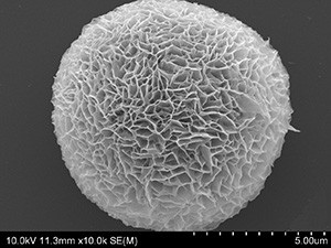
'Microflower'

SEM image of a synthesized organic-inorganic microflower structure.

Imaged on the Hitachi SU-70 scanning electron microscope, at Auckland University of Technology (AUT)

Shanshan was not able to receive her award in person at the celebration but was presented with it by a BIRU staff member later.

See Shanshan with her prize

Visualisation and analysis


Anandita-300px

Anandita Umapathy

Department of Obstetrics and Gynaecology, School of Medicine, FMHS

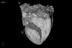
'What becomes of the broken hearted'

An E18.5 mouse fetal heart that “cracked” whilst undergoing chemical air drying to visualise cardiac fibre patterns.

It was imaged using microCT (Bruker SkyScan 1272) and visualised using CTVox software (Bruker) at the Auckland Bioengineering Institute.

Acknowledgements: Mr. Dane Gerneke (ABI)

See Anandita with her prize