Biomedical Imaging Research Unit

Image competition results - 2023

The award winners of the 2023 BIRU image competition were announced on Tuesday 28 November at the annual BIRU Research Celebration. As usual, there was a very high standard among the competition entries. The category winners are shown below. The highly commended award winners can be viewed here.

Click on each image to see a larger version.

Visualisation and analysis and trophy winners for 2023


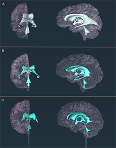
3-VS-CP-BIRU comp image_300px

Vivi Sheppard and Caleb Paterson

School of Medical Sciences

'Morphological Changes of the Aging Human Cerebroventricular System'

Cerebral ventricular system - a cavity where cerebrospinal fluid is produced, acting mainly as a mechanical shock absorber for the brain and the spinal cord and as a part of the waste clearance system for the brain, known as the glymphatic system.

Software used for 3D visualisation and segmentation of MR images - Amira

See Caleb with the prizes here.

Electron microscopy


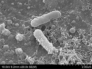
EM_2023_300px

Saurabh Lamba

School of Chemical Sciences

'A Bursting Tale of E.coli'

This tale is of E. coli attempting to colonize a urinary catheter and forming a biofilm. However, little does E. coli know that the catheter is coated with a protective layer of antimicrobial lipopeptide, which kills the bacteria as it comes in contact with the catheter surface.

Imaged on the FEI Quanta 200 F ESEM microscope at 10 kV, at Auckland University of Technology (AUT)

Confocal microscopy


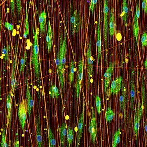
CLSM_2023_300px

Amy McCaughey-Chapman

Pharmacology & Clinical Pharmacology/Centre for Brain Research

'Christmas lights'

Human reprogrammed oligodendrocytes (green) cultured on nanofibers. The oligodendrocytes are stained with the marker O1.

Image was captured on the Zeiss LSM 710 confocal microscope using the Plan Apochromat 20x/0.8 NA objective.
 

Light microscopy


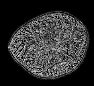
LM_2023_300px

Dr Charlotte (Charlie) Johnson

School of Biological Sciences

Stitched image of a teardrop, allowed to dry and crystallise.

Captured using a Nikon Ti-E inverted microscope at 4x magnification using DIC/polarisation with an Andor Clara camera.

Stitched and edited in Adobe Photoshop.