Biomedical Imaging Research Unit

Highly commended and special awards - 2016

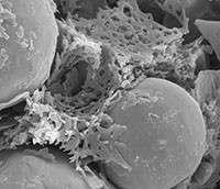
Electron microscopy - highly commended


19 - Nurual -4-200px

Nurul Haiza Sapiee -Chemical & Materials Engineering

Polymethyl methacrylate (PMMA) binding titanium powder to aid injection moulding process

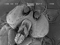
Electron microscopy - highly commended


08 -Bianca Heilman_Image5-200px

Bianca Heilman – Department of Physiology

SEM of the head of a fruit fly (Drosophila melanogaster)
 

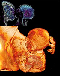
Visualisation and analysis - highly commended


08- Amirjalili - The Life-200px

Ali Mirjalili - Department of Anatomy with Medical Imaging

“The Life” - MRI of a baby during breast feeding

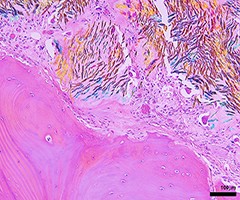
Light microscopy - highly commended


03 -Ashika Chhana 1_Gouty bone erosion in human joint_polarised light-200px

Ashika Channa - Department of Medicine

Bone erosion in a human joint tissue affected by gout.

Stained with H&E (by Satya Amirapu), viewed using polarising light microscopy with a red filter. Uric acid crystals (the causative agent of gout) are orange and blue under these settings. The image shows inflammatory cells and uric acid crystals adjacent to bone.

See Ashika receiving her prize from Sue
 

Light microscopy - special mention


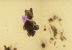
21- Alyona -2_14.57.13-200px

Olena Oryshchuk  - Department of Molecular Medicine and Pathology

Captured on the Nikon Microscope !!

"Finding this teddy bear gave me so much joy in a stressful time of my Honours year".  

Confocal microscopy - highly commended


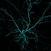
03 -Wojciech A - hippocampal neuron MAP2-200px

Wojciech Ambroziak – Department of Physiology/Centre for Brain Research

This image represents a hippocampal pyramidal neuron labelled for MAP2 acquired with ZEISS LSM 710 Inverted Confocal Microscope

Visualisation and analysis - highly commended


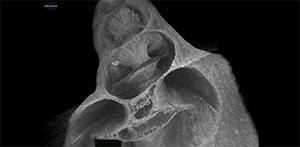
Matthew-Cochlea-300px

Matthew Fields - Auckland Bioengineering Institute
“Cochlea Model.bmp” is a reconstructed 3D model of a guinea pig cochlea after staining with osmium tetroxide and imaging using microCT
 

Light microscopy - highly commended


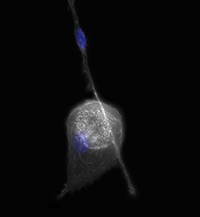
26 - JMathy-LM1-200px

Joanna Mathy - School of Biological Sciences

Two Ships – This image captures the two predominant, morphologically-distinct cell types that differentiate from human peripheral blood monocytes when grown in tissue culture: round and spindle

Ships that pass in the night, and speak each other in passing,
Only a signal shown and a distant voice in the darkness;
So on the ocean of life we pass and speak one another,
Only a look and a voice, then darkness again and a silence.

1874, Henry Wadsworth Longfellow, Tales of a Wayside Inn
 

See Joanna receiving her prize from Hilary

Confocal microscopy - highly commended


25 - nakazawa-2-200px

Yosuke Nakazawa – Department of Physiology

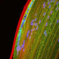
Mouse Lens axial sectioning image labelled with TRPV4, WGA, and DAPI

See Yosuke receiving his prize from Hilary