Dr Clairton de Souza
Department of Ophthalmology
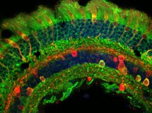
Vertical section of Human retina
Acquired on the Olympus FV1000 confocal microscope in the Department of Ophthalmology
Click on thumbnail image to see larger version.
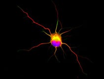
Rat hippocampal neuron grown in culture for 2 days. Blue is a DAPI stain of DNA in the cell showing the cell nucleus, while red is an immunocytochemical stain for acetylated tubulin (a stable form of the protein) and green is for tyrosinated tubulin (unstable form of the protein).
Imaged using a Leica DMR microscope with a SPOT pursuit-XS camera in the School of Biological Sciences.
Click on thumbnail image to see larger version.
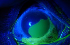
Fluorescein dye in a human eye showing epithelial recovery after a chemical burn and treatment with connexin43 specific antisense oligonucleotides. The dye (green) adheres to connective tissue where the epithelium has not yet recovered.
Click on thumbnail image to see larger version.
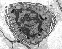
Mast cells
Imaged on the BIRU Tecnai G2 Spirit TWIN electron microscope.
Click on thumbnail image to see larger version.
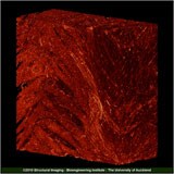
3D reconstruction of a section of the left ventricle of rat heart 35 days after a myocardial infarct.
Imaged on the Large Volume Imaging System in the Department of Physiology.
Click on thumbnail image to see larger version.
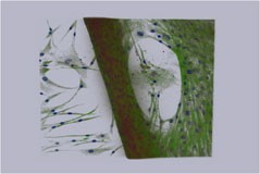
“Tidal wave” This extensive structure was formed directly in vitro from cells isolated from the cornea. This structure is reminiscent of the limbus of the eye where the clear cornea and the white sclera meet. This tidal wave-like structure is morphologically distinct and retains the labelling of neural precursors which has been demonstrated in limbal stem cells. Labelled for Musashi-1 (Alexa568, red), nestin (Alexa488, green) and nuclear counterstain with DAPI (blue).
Image data acquired on the Olympus Fluoview 1000 confocal microscope in the Department of Ophthalmology.
Volume rendering and movie was generated using the AMIRA software.
Click on thumbnail image to see larger version.

