Biomedical Imaging Research Unit

Image competition results - 2014

Visualisation and Analysis category and trophy winner


Inken_TrophyImage_LymphNode_160px

Inken Kelch

School of Biological Sciences

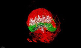
3D reconstruction of a mouse lymph node, extended-volume confocal imaging system

Image processing was performed using custom-developed computer tools, the 3D rendering software Amira, and Imaris was used for the creation of this animation.

Link to publication

Click on the thumbnail image to see a larger version.

See Inken with her prizes

View another V&A entry that received a highly commended award

Light microscopy winner - joint equal


Zakieh_Light_160px

Zakieh Vahdati

Infection and Immunity, School of Medical Sciences

Supervisors - Dr Siouxsie Wiles and Dr Simon Swift

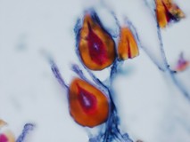
A beautiful structure in outer membrane of the cuticle ( probably spiracles) in Galleria Mellonella larvae. Light microscopy, acid fast staining.

Click on the thumbnail image to see a larger version.

See Zakieh with her prizes

View another Light Microscopy entry that received a highly commended award
Light microscopy winner - joint equal


RaviSharon_Light_px

Ravi Jain and Sharon Waldvogel

Department of Surgery, FMHS

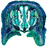
Coronal section through the snout and paranasal sinuses of a germ free mouse (10-12 weeks of age).

Stain: Sirius red-light green.

Click on the thumbnail image to see a larger version.

See Sharon with her prizes

View another Light Microscopy entry that received a highly commended award
 

Confocal microscopy winner


Renita_Winner_Confocal_160px

Renita Martis

Department of Optometry and Vision Science

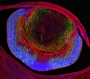
An axial image of the embryonic eye (e16) labelled with cystine/glutamate antibodies, WGA and DAPI

Acquired using the Olympus FV1000 confocal microscope
Click on the thumbnail image to see a larger version.

See Renita with her prizes

View another Confocal Microscopy entry that received a highly commended award