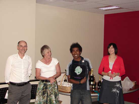
School of Medical Sciences


2008 competition - winning entries

Visualisation and analysis winner and trophy winner


Overrall winner

Isuru Dilshan
Biophysics and Biophotonics Laboratory
Department of Physiology
School of Medical Sciences

A reconstructed slice through a cardiac muscle cell. More details

Imaged on the Olympus FV1000 confocal microscope.

Click on thumbnail image to see larger version. See Isuru receiving his prize.

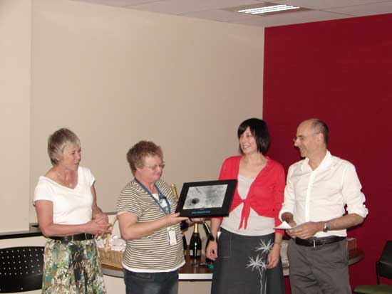
View the  Highly commended image

Confocal laser scanning microscopy winner


vithika_jinny_winner_clsm_160px

Vithika Suri and Jinny Yoon,
Department of Anatomy with Radiology
School of Medical Sciences

A whole-mount section of an organotypic hippocampal slice cultured for 14 days. Shown in the image is the CA3 area of the hippocampus.
Green (Alexa488) represents Connexin43 (gap junction protein), the red (Alexa568) represents MAP2 (neuronal marker), and the blue (Cy5) represents glial fibrilary astrocytic protein (astrocytic marker).

Imaged on the Liggins Zeiss LSM 510 META confocal microscope.

Click on thumbnail image to see larger version. See Jinny receiving her prize on behalf of herself and Vithika.

Light microscopy winner


jchua_winner_lm_160px

Dr Jacqueline Chua
Department of Optometry and Vision Science
Faculty of Science

Amino acid staining through mouse retinal cells. The red channel is positive for glutamate, green channel for GABA and blue channel for glycine. The mouse tissue was cut horizontally and thinly focusing on the inner nuclear layers.

Imaged on the BIRU's Leica DMR light microscope.

See Jacqueline receiving her prize.

View the three Highly commended images.

Transmission electron microscopy winner


lindagraham_winner_tem_160px

Linda Graham
LabPLUS

Human skeletal muscle

Accidentally taken while changing magnifications! Hence double or it may even be triple exposure.

Imaged on the BIRU's Tecnai G2 Spirit TWIN TEM.

See Linda receiving her prize.