Biomedical Imaging Research Unit


Image competition results - 2013

Visualisation and Analysis category and trophy winner


2013_va_trophywinner_160px

Corin Gardiner

School of Biological Sciences

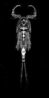
Orthographic projection of the carapace of a Cyclopoid copepod. Ventral view

The image was acquired on the Zeiss LSM 710 confocal microscope and rendered using Amira.

Click on the thumbnail image to see a larger version.

See Corin with his prizes

View another V&A entry that received a highly commended award

Confocal microscopy winner


2013_clsm_winner_kj_160px

Kathryn Jones
Neural Repair and Neurogenesis Group, Department of Pharmacology & Clinical Pharmacology/Centre for Brain Research


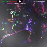
Human skin cells are transformed into functional neurons.


This image shows induced neural progenitor cells (yellow) and immature neurons (blue) that have been reprogrammed from human skin.

Imaged on the Zeiss LSM 710 confocal microscope.

Click on the thumbnail image to see a larger version.

See Amelia collecting the prize on behalf of Kat.

View other confocal microscopy images that received awards

Electron microscopy winner


2013_tem_winner_lg_160px

Linda Graham

LabPlus

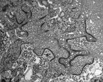
Terminal Junction = renal proximal tubule junctional complexes

Imaged on the Tecnai G2 Spirit Twin electron microscope.

Click on the thumbnail image to see a larger version.

See Linda receiving her prize.


View another electron microscope image that received a special mention award

Light microscopy winner


2013_lm_winner_si_160px

Salim Ismail, Jane McGhee, Trevor Sherwin
Department of Ophthalmology

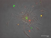
Show your true colours.

FUCCI (Fluorescence Ubiquitination Cell Cycle Indicator) transduced corneal cells indicate their stage of the cell cycle as they migrate out from a central sphere. Red indicates G1 of the cell cycle, Green indicates S/G2/M stages of the cell cycle and yellow indicates cells in the G1 to S transition. Imaged using the FLOID Cell Imaging Station, fluorescence channel images overlaid using Adobe Photoshop

Equipment: FLOID Cell Imaging Station; Adobe Photoshop

See Salim receiving his prize.

View other light microscope images that received highly commended awards