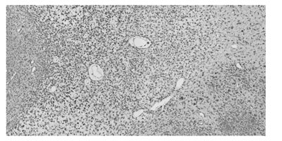
Biomedical Imaging Research Unit

Highly commended and special awards - 2020


Confocal microscopy - highly commended


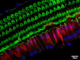
CLSM_HC_2020_200px

Ziyin (Silver) Huang

Department of Physiology/Audiology

“These cells listen for you”

Labeling shows expression of P2X4, stereocilia and nucleus on one row of inner hair cells and three rows of outer hair cells in cochlea. Image acquired on BIRU ZEISS LSM 800 Airyscan

Electron microscopy - highly commended


EM_HC_2020_200px

Urawadee Rajchakit and Hugh Glossop

School of Chemical Sciences

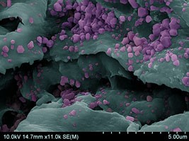
“Gold mine”

Hexagonal-shaped gold nanoplates embedded in peptide fibers for water purification application.

SEM gold nanoplates coloured using Photoshop.

Image acquired using the Hitachi SU-70 FESEM at the Auckland University of Technology (AUT).

Visualisation and analysis - highly commended


VA_HC_2020_200px

Hanna Allerkamp

Department of Obstetrics and Gynaecology & Auckland Bioengineering Institute

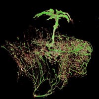
“The tree of life”

Utero-placental arteries of a rat, injected with a contrast agent, imaged by microCT (SKYSCAN 1272, Bruker) and finally visualized and processed with the CTVOX software (Bruker) at the ABI

Acknowledgements:

Mr Dane Gerneke (ABI)

See Hanna receiving her prize from BIRU Director Dr Gus Grey.

Light microscopy - highly commended


LM_HC2_2020_200px

Saem Park

School of Biological Sciences

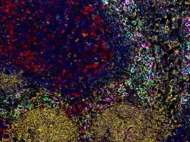
“Painting tumour tissue”

Melanoma-infiltrated lymph node stained with a 7-colour immune panel. Images were acquired on the Vectra Polaris at 20x magnification.

See Saem receiving her prize from BIRU Director Dr Gus Grey.

LM_HC1_2020_200px

Scott Bolam

Department of Surgery

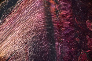
“Entheseal Entwinement”

Murine supraspinatus tendon enthesis imaged on a Nikon Ni-E Microscope under polarised light with x10 magnification.

Light microscopy - special mention


LM_SM_2020_200px

Kelly Zhou

Department of Physiology

"Smiley face"

Blood vessels forming the outline of a smiley face in a white matter tract of the brain. Taken on BIRU MetaSystems VSlide slide-scanner.