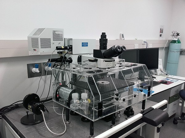
- LASERS:
Argon ion multi-line (458nm, 488nm, 514nm)
Helium Neon: 543nm, 633nm
Diode-pumped 405nm (for imaging and photoactivation/bleaching) - Twin scanner system. SIM scanner for simultaneous bleaching and imaging capability
- Detectors: 2 spectral (variable bandpass), 1 filter-based, and one transmitted light + 2 high sensitivity detectors (HSD) for green/red and red/far-red
- Spectral unmixing
- Modes: xy, xyz, xt, xyt, lambda
- Resolution: up to 4096 x 4096 pixel resolution
- Filter blocks for viewing specimens: DAPI, FITC, TRITC, CFP
- Four separate nosepieces, one for water-immersion long working distance objectives, one for the 20x high NA water immersion lens, one for the 10x Scaleview lens and one for dry and oil immersion objectives (for imaging slides)
- Objectives: long working distance water-immersion (10x/0.3 NA, 20x/0.5 NA, 40x/ 0.8 NA, 60x/ 0.9 NA), dry (4x/0.16 NA, 10x/0.4 NA, 20x/0.8 NA, 40x/0.95 NA), silicone oil immersion (60x/ 1.3 NA) and oil immersion (50x, 60x/ 1.35NA, 100x/1.4 NA)
Specialised 10x/0.6 NA XPLN SVMP lens, larger circumference than other lenses, working distance 8mm, refractive index (RI) immersion capability from 1.33-1.52
20x/1.0 NA long working distance water-immersion lens - larger circumference than standard water-immersion lenses - Solent incubation system with heating and 5% carbon dioxide supply
- Kinetic Systems anti-vibration table


