-
Biomedical Imaging Research Unit
- Our unit
- Microscopy and imaging
- Tissue processing
- Flow cytometry
- Image processing and analysis
-
BIRU image competition
- 2014 trophy winner
- 2013 trophy winner
- 2013 winners
- 2013 commended
- 2012 trophy winner
- 2012 winners
- 2012 commended
- 2012 special award
- 2011 trophy winner
- 2011 winners
- 2011 commended
- 2011 special awards
- 2010 trophy winner
- 2010 winners
- 2010 commended
- 2009 trophy winner
- 2009 winners
- 2009 commended
- 2008 trophy winner
- 2008 winners
- 2008 commended
- 2007 winners
- 2007 commended
- 2006 winners
- 2006 commended
- 2014 winners
- 2014 commended
- 2015 winners
- 2015 commended
- 2015 trophy winner
- 2015 special mention
- 2016 winners
- 2016 commended
- 2017 winnners
- 2017 commended
- 2018 commended
- 2018 winners
- 2019 winners
- 2019 commended
- 2019 trophy winner
- 2020 winners
- 2020 commended
- 2020 trophy winner
- 2021 trophy winner
- 2021 winners
- 2021 commended
- 2021 special award
- 2022 winners
- 2022 commended
- 2022 trophy winner
- 2022 special awards
- 2023 winners
- 2023 commended
- 2024 winners
- 2024 commended
- 2024 special mention
- 2025 winners
- 2025 commended
- Event photos
- Resources and links
- Teaching and learning
- Our people
- Equipment charges
- Seminars and workshops
University home
parent of
Faculty of Medical and Health Sciences
parent of
School of Medical Sciences
parent of
ABOUT
parent of
Our departments
parent of
Biomedical Imaging Research Unit
parent of
BIRU image competition
parent of
2020 trophy winner
Biomedical Imaging Research Unit
2020 Hilary Holloway Prize for best image - Joscelin Smith
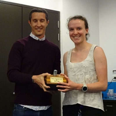
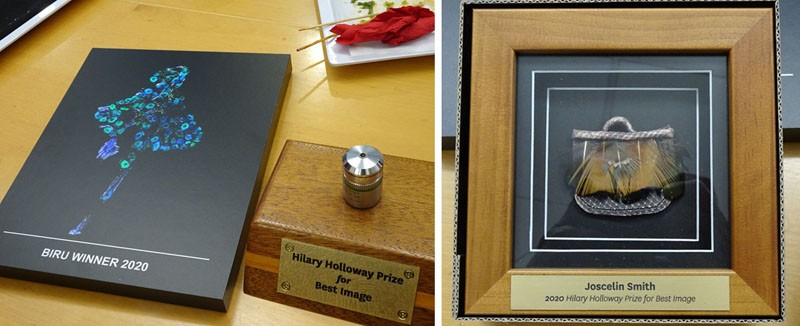
Joscelin Smith, from the Department of Physiology is the 2020 winner of the Hilary Holloway Prize for best image. Joscelin was the winner of the Confocal Microscopy category. In addition to keeping the trophy for one year, Joscelin received a special "Basket of Knowledge" award. The photo below shows Joscelin receiving her award from BIRU Director Dr Gus Grey.
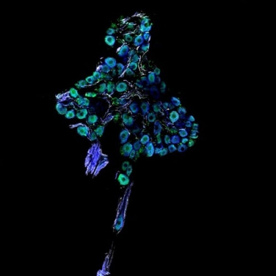
The winning image is shown below (click on the image to see a larger version). Entitled "Deep sea neurons", the image shows ganglionated plexi neurons in the heart. It was stained for neuron specific markers (GluR2, ChAT and NFH and acquired on the Olympus FV1000 confocal microscope using a 10x/0.4 NA objective lens.
-
Biomedical Imaging Research Unit
- Our unit
- Microscopy and imaging
- Tissue processing
- Flow cytometry
- Image processing and analysis
-
BIRU image competition
- 2014 trophy winner
- 2013 trophy winner
- 2013 winners
- 2013 commended
- 2012 trophy winner
- 2012 winners
- 2012 commended
- 2012 special award
- 2011 trophy winner
- 2011 winners
- 2011 commended
- 2011 special awards
- 2010 trophy winner
- 2010 winners
- 2010 commended
- 2009 trophy winner
- 2009 winners
- 2009 commended
- 2008 trophy winner
- 2008 winners
- 2008 commended
- 2007 winners
- 2007 commended
- 2006 winners
- 2006 commended
- 2014 winners
- 2014 commended
- 2015 winners
- 2015 commended
- 2015 trophy winner
- 2015 special mention
- 2016 winners
- 2016 commended
- 2017 winnners
- 2017 commended
- 2018 commended
- 2018 winners
- 2019 winners
- 2019 commended
- 2019 trophy winner
- 2020 winners
- 2020 commended
- 2020 trophy winner
- 2021 trophy winner
- 2021 winners
- 2021 commended
- 2021 special award
- 2022 winners
- 2022 commended
- 2022 trophy winner
- 2022 special awards
- 2023 winners
- 2023 commended
- 2024 winners
- 2024 commended
- 2024 special mention
- 2025 winners
- 2025 commended
- Event photos
- Resources and links
- Teaching and learning
- Our people
- Equipment charges
- Seminars and workshops
-
SCHOOLS, DEPARTMENTS AND CENTRES
Connect with us
Connect with us
-
SCHOOLS, DEPARTMENTS AND CENTRES


