Biomedical Imaging Research Unit

Highly commended, special and patriot awards - 2017

The following awards were presented at the BIRU end of year Research Celebration on Thursday 23 November 2017. Congratulations to all of those who received awards and thanks to all who participated this year.

Light microscopy - highly commended


YuktiVyas_small

Yukti Vyas
Centre for Brain Research / Department of Physiology

Patching Dendrite and Cell body of cortical neuron: cortical neuron patched (using electrophysiology) at the cell body and dendrite 384um apart, and filled with fluorescent dye to visualise this neuron amongst all the others in the cortex (imaged at 40x magnification).

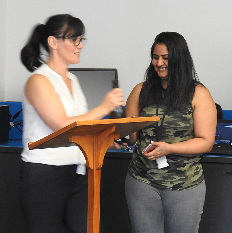
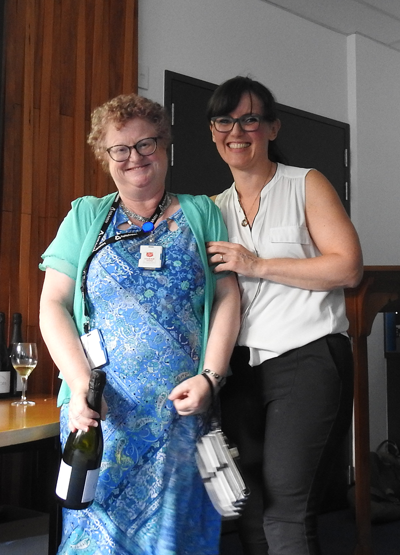
See Yukti receiving her prize from BIRU Director Dr Sue McGlashan!
 

Light microscopy - highly commended


KathrynJones_small

Kathryn Jones

School of Biological Sciences

“Cell reprogramming – transforming skin into brain”
Human neurons with long processes connecting up and communicating with each other (TuJ1 - green), while expressing a dopamine generating enzyme called AADC (red). Cell nuclei are labelled in blue. These beautiful cells were actually mature human skin only four months earlier and reprogrammed into pluripotent stem cells, before being directed down the neuronal lineage and matured into neurons in the lab.
 

Light microscopy- special mention


AmandaCharlton_small

Amanda Charlton

LabPlus

Micro Array of Macroscopic Animalcules

Microarrays are multiple 2-3mm cores of tissue assembled into a single paraffin block commonly used in tissue research to enable comparison between different specimens on a single glass microscope slide.
 

Electron microscopy - highly commended


LindaGraham_small

Linda Graham
LabPlus, ADHB

" Snow Flake series"

Stain precipitates, probably Uranium

See Linda receiving her prize from BIRU Director Dr Sue McGlashan!
 

Confocal microscopy - highly commended


ElishaHayden_small

Elisha Hayden

Department of Molecular Medicine and Pathology

The Killer Egg

Formation of a 3D structure arising naturally from a 2D co-culture composed of melanoma cells, stably expressing GFP, with human dermal fibroblasts, stained red for CD90. Imaged on Zeiss LSM 710 confocal microscope.

See Elisha receiving his prize from BIRU Director Dr Sue McGlashan!
 

Visualisation and analysis - highly commended


ChanJin_small

Chan Jin

Department of Physiology

Three-dimensional volume rendering of a guinea pig head acquired using micro-computed tomography (SkyScan 1172, Bruker). Some images have been sliced along the longitudinal axis of the cochlea (posteriorly-located spiral-shaped component of the inner ear) for display purposes.

Visualisation and analysis - highly commended


EmmaKang_small

Emma Kang

School of Pharmacy

A brain tumour cell image taken in BIRU using the Nanolive 3D Cell Explorer.

Visualisation and analysis - special mention


Miriam Scadeng_+small

Miriam Scadeng

Department of Anatomy and Medical Imaging

A spiral acquisition on an anaesthetized emperor penguin using a Toshiba clinical CT scanner. Air sacs of the respiratory system of the Emperor penguin - in vivo. Organ segmentation was done with Amira software.
 

Visualisation and analysis - Patriot award


MonicaAcosta_small

Monica Acosta
School of Optometry and Vision Science

“Incomplete sagittal MRI scan of a New Zealander head”