Linda Graham
LabPlus
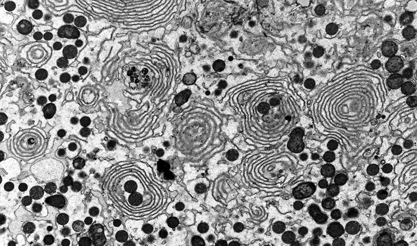
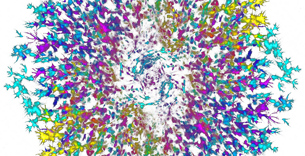
Rough Endoplasmic Reticulum


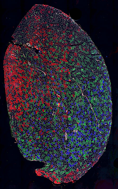
Rat skeletal muscle
Channels: red-Type IIb fibres, I green- type IIa fibres, blue- type I fibres, White- membrane of muscle fibres (sarcolemma). Then unstained in black is type IIx fibres.
Acquired on the MetaSystems VSlide microscope.

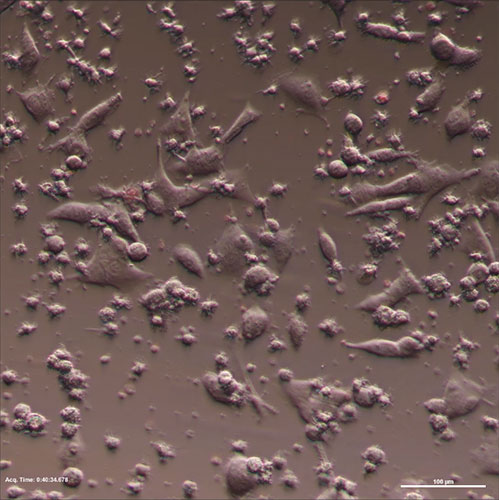
A melanoma cell line (Trombelli) getting killed by a T cell clone (1D4).

Maximum projection – modified using Photoshop
Replaced black with white, mirrored the image with some careful blending of the mirrored layer, and then adjusted the hue of the whole image.

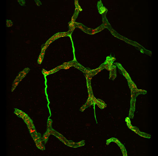
String vessels
Narrowed blood vessels with empty basement membrane tubes (green) and no endothelium (red), in frontal cortex of human brain with Parkinson disease.

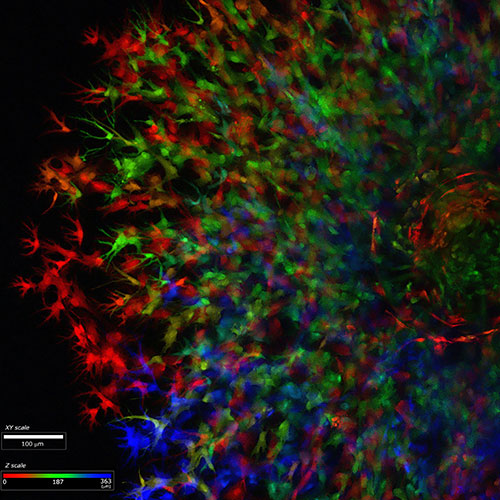
Maximum projection, which has been colour coded for depth in ZEN software.
The injection site is visible as a kind of ring shape on the right-hand side of the image. Colour coding for depth allows visualisation of the extent of cell migration at different depths through the gel by looking at a single 2D image, rather than having to create a movie.
