Ratish Kurian
IT Systems Specialist - BIRU
Email: r.kurian@auckland.ac.nz
Phone: +64 (0) 9 923 6195
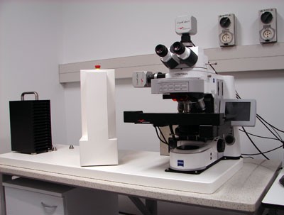
CoolCube1 (colour) CCD
CoolCube 4m TEC (monochrome) sCMOS
Camera upgrade funded by the Liggins Institute in 2019
Solid-State Light Source Colibri 7
7 solid state LED lamps - wavelengths below
UV (385nm) for excitation of DAPI, Alexa 405, Hoechst 33258 and similar dyes
Violet (430nm) for excitation of eCFP, Lucifer Yellow, Alexa 430 and similar dyes
Blue (475nm) for excitation of eGFP, Fluo4, FITC and similar dyes
Green (555nm) for excitation of Cy3, TRITC, DsRed and similar dyes
Yellow (590nm) for excitation of mCherry, Alexa 568, mPlum and similar dyes
Red (630nm) for excitation of Cy5, Alexa 631, TOTO-3 and similar dyes
Far Red (735nm) for excitation of Cy7 and similar dyes
Band width details available here

Multiband filter sets
81 HE – QUAD filter, suitable for fluorescent dye combinations like DAPI, FITC, DsRed, Cy5
Uses LEDs 385, 475, 555, 630nm
Collects emission bands 375/38 + 484/25 + 553/18 + 631/22
Spectra here
112HE – PENTA filter, suitable for fluorescent dye combinations like DAPI, FITC, DsRed, Cy5 and Cy7
Uses LEDs 385, 475, 555, 630, 735nm
Collects emission bands 425/30 + 514/31 + 592/25 + 681/45 + 785/38
Spectra here
Single band filter sets
Filter set #01 (Use LED 385; Em- 447/60nm) - spectra
Filter set #02 (Use LED 430; Em-550/32nm) - spectra
Filter set #03 (Use LED 475; Em 527/20nm) - spectra
Filter set #04 (Use LED 555nm; Em 580/23nm) - spectra
Filter set #05 (Use LED 590; Em 628/32) - spectra
Filter set #06 (Use LED 630; Em 676/29nm) - spectra
Filter set #07 (Use LED 735; Em 809/81nm) - spectra
If you want to see some additional examples which illustrate what can be done with this system, please take a look at the movies below which were created by Luke Hammond from QBI Microscopy using his VSlide.

ImageJ macros for cropping borders from extracted images:


Successful grant application by Professor Mike Dragunow (Dept of Pharmacology & Centre for Brain Research) to Gravida: National Centre for Growth and Development
The system was upgraded with a CoolCube 4mTEC monochrome sCMOS camera funded by the Liggins Institute in 2019
Subscription rate: $52.00 per hour (10 hours minimum) – pay in advance
Casual rate: $61.73 per hour – pay as you go

Ratish Kurian
IT Systems Specialist - BIRU
Email: r.kurian@auckland.ac.nz
Phone: +64 (0) 9 923 6195
