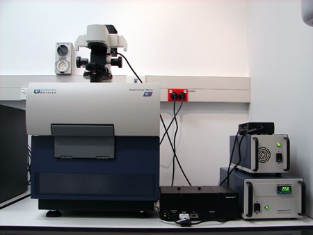
- Fast automated acquisition
- Automated customised analysis routines
- Hardware (laser-based) autofocus capability
- Software (image-based) focusing capability
- Multi-well plates up to 1536 wells
- Chamber slides
- Fluorescence imaging
- Brightfield imaging
- Phase contrast imaging (20x only)
- Live cell imaging - multi-site, multi-wavelength
- Z series
- Tile-scanning
Biomedical Imaging Research Unit
Molecular Devices ImageXpress Micro XLS
High content screening system

Specifications
Camera - Andor Zyla CMOS
Objective lenses
- 2x/0.1 NA CFI Plan Apochromat
- 4x/0.2 NA S Fluor
- 10x/0.3 NA Plan Fluor
- 20x/0.45 NA CFI Super Plan Fluor ELWD ADM (phase contrast)
- 40x/0.6 NA S Plan Fluor ELWD
Three slide holder - for chamber slides
Environmental control - heating and carbon dioxide gas supply
Light sources
- Lumencor Spectra X configurable light engine - fluorescence imaging (filter specifications below)
- 100 W Halogen lamp - transmitted light
Excitation and emission filters

List of excitation and emission filters for Molecular Devices ImageXpress High Content Screening system (69.9 kB, PDF)
Software and application modules
MetaXpress, Custom Module Editor, Autoquant deconvolution
Application modules - Angiogenesis • Cell Cycle • Cell Health • Live/Dead • Multiwavelength Cell Scoring • Neurite Outgrowth • Transfluor • Translocation • Multi Wavelength Translocation • Micronuclei • Granularity
Documentation from Molecular Devices is available for University of Auckland staff/students here. Please note that this is a secure page and these files should not be shared without permission from a BIRU staff member.
Specimen requirements
The IXM microscope stage requires plates to have a base footprint of 128mm x 86mm, with at least 3 square corners present. Please check that your plate meets these requirements.
Please note - the NUNC 142475 24 well plate and the NUNC 150628 12 well plate do not fit the stage so cannot be used as three of the corners are cut off.
Contact person
Ratish Kurian
Senior Technologist/IT Systems Specialist - BIRU
Email: r.kurian@auckland.ac.nz
Phone: +64 (0) 9 923 6195
-
SCHOOLS, DEPARTMENTS AND CENTRES
Connect with us
Connect with us
-
SCHOOLS, DEPARTMENTS AND CENTRES

