Biomedical Imaging Research Unit

Light sheet fluorescence microscopy comes to BIRU

09 December 2024

Light sheet fluorescence microscopes work by using a sheet of light for excitation rather than a spot/point as per point-scanning confocal microscopes. They are designed for speed and particularly useful for model organisms that are already  transparent such as zebrafish embryos, C. elegans, etc. as well as  thicker optically cleared samples such as tissue blocks, spheroids/organoids. The resolution of these systems sits between widefield fluorescence and confocal microscopy and there is a wide variety of designs available for these systems.

The system was funded through a successful application to The University of Auckland Shared Research Infrastructure fund in 2023. A working group was tasked with the job of deciding on the most suitable system for the BIRU. The group was led by Jacqui Ross on behalf of BIRU with 6 other staff members; Dr Haruna Suzuki-Kerr/Physiology, Dr Jiwon Hong/SBS, Associate Professor Chris Hall/MMP, Associate Professor Jo James/O&G, Dr Rashi Karunasinghe/Physiology and Dr David Crossman/BIRU Director/Physiology.

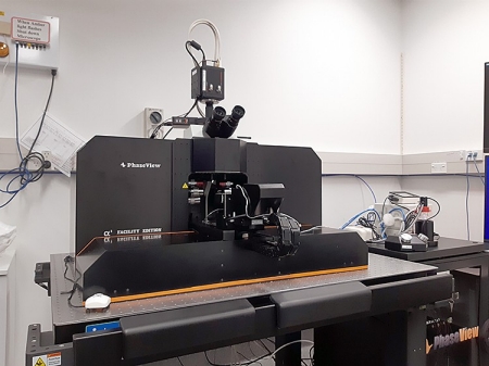
PhaseViewAlpha3LightSheet_750px

The system selected by the team was a PhaseView Alpha 3 Facility Edition light sheet fluorescence microscope. It was recently in stalled in BIRU and has a wide variety of objective lenses. There are 6 excitation lasers and 6 separate filter sets. An Okolab incubation system is available that can provide heating and 5% carbon dioxide.

This microscope is designed for core facilities and most functions are motorised for ease of use. Calibration using the preferred immersion medium is required in advance of using the system in order to align the light sheet arms. The software is quite straightforward to use and Imaris is available for advanced visualisation and quantification. A separate high specification PC is available for this purpose.

For a full list of specifications, please check out the system page.

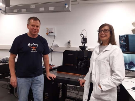
PhaseViewLightSheetMicroscope_JR_IL
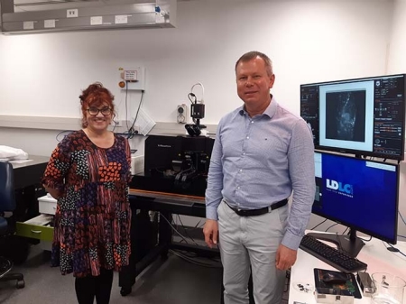
Igor Lyuboshenko (PhaseView) and Jacqui Ross (BIRU) after installation of the new system
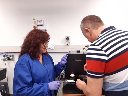
PhaseViewLightSheetMicroscope_IL_SR
Sandrine Roy (Evident Scientific) and Igor Lyuboshenko (PhaseView) get a sample ready for testing the system
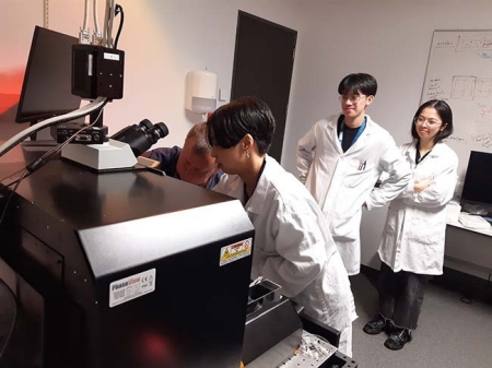
PhaseViewLightSheetMicroscope_HS
Haruna Suzuki-Kerr (Physiology) and her students test out the new system with their sample with help from Igor Lyuboshenko (PhaseView)
PhaseViewLightSheetMicroscope_SR_IL
Sandrine Roy (Evident Scientific) and Igor Lyuboshenko (PhaseView) after installing the new system